Podcast: Play in new window | Download (Duration: 49:44 — 69.2MB)
Subscribe: RSS
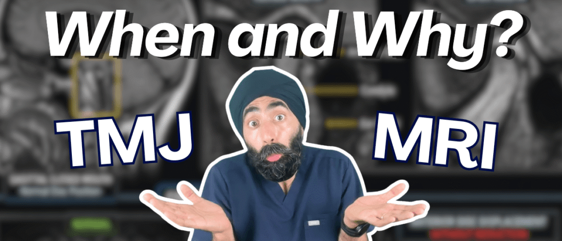
When is it appropriate to consider an MRI for your TMD patient?
What’s actually involved in MRI of the TMJ?
Can you use any MRI machine, or is the choice of imaging center crucial?
And who should be reporting on these scans — does it really matter? (Hint: yes, it does!)
Dr. Kevin Lotzof, a straight-talking radiologist, joins Jaz for a controversial deep dive into the role of MRI in Temporomandibular Disorders. While many experts downplay its importance, Kevin argues that TMJs are under-imaged and under-diagnosed — and that we may be missing critical pathology.
They explore the practicalities of imaging, how to set expectations with your patients, and why strong but differing views in TMD care can ultimately help you refine your own clinical approach.
Protrusive Dental Pearl: Adopt the mindset of “Find the cancer today.”
When carrying out examinations—whether soft tissue or extraoral—approach it with the intention of detecting oral or skin cancers early. This mindset helps clinicians look beyond just teeth, catch unusual or suspicious lesions, and potentially save lives.
Key Takeaways
- TMJ is often overlooked but is crucial for overall health.
- MRI is essential for accurate TMJ diagnosis.
- Cone beam CT cannot replace MRI for TMD assessment.
- Patients with headaches may have undiagnosed TMD.
- Education on TMJ imaging is lacking among dental professionals.
- Asymptomatic patients should still be scanned for TMJ issues.
- The quality of imaging directly impacts diagnosis accuracy.
- Patients often feel anxious about MRI procedures.
- Understanding patient perspectives can improve care.
- There is a need for better collaboration between dentists and radiologists.
Highlight of the episode:
- 00:00 Teaser
- 00:55 Intro
- 05:20 Protrusive dental pearl
- 06:36 Interview with Dr. Kevin Lotzof
- 09:38 Under-Imaging and Differing Perspectives
- 13:27 Access and MRI Centers in the UK
- 17:51 TMJ MRI: Patient Expectations
- 22:17 Midroll
- 25:53 Open MRI Machines
- 27:26 Ideal Candidates for MRI Imaging
- 29:55 Cone Beam CT vs. MRI
- 31:53 Screening and Asymptomatic Patients
- 38:43 Centers with Reliable TMJ Imaging
- 41:27 Encouragement for General Dentists
- 46:33 Outro
Where to Get Reliable TMJ Imaging
⭐ Top Pick:
- Orion, Wimpole Street, London
(Full contact details available via the Protrusive Guidance App)
🏙️ Other London Options:
- Spire Bushey, Circle Hendon, Cavell, Kings Oak, Circle Healthcare Center
Learn more about TMJ radiographic imaging in PDP223: Understanding TMD Radiographic Imaging – Pano vs CBCT vs MRI
#PDPMainEpisodes #OcclusionTMDandSplints #CareerDevelopment
This episode is eligible for 0.75 CE credit via the quiz on Protrusive Guidance.
This episode meets GDC Outcomes A and C.
AGD Subject Code: 730 ORAL MEDICINE, ORAL DIAGNOSIS, ORAL PATHOLOGY (Imaging techniques)
Aim: To highlight the importance of MRI in the diagnosis and management of temporomandibular joint (TMJ) disorders, ensuring safe and effective orthodontic and restorative treatment planning.
Dentists will be able to:
- Explain why MRI is superior to clinical examination and CBCT in diagnosing TMJ pathology.
- Identify the key indications for TMJ MRI, including both dental and non-dental symptoms.
- Recognize the limitations of poor imaging technique and reporting in TMJ diagnosis
Continuing Education Information
This activity may be eligible for continuing education credit through Protrusive Guidance. Participants must complete the associated quiz inside Protrusive Guidance to obtain a CPD/CE certificate.
Cost:
Access to this CE activity is included with an active Protrusive Guidance membership. Current membership pricing is available at www.protrusive.app.
Cancellation & Refund Policy:
Memberships may be cancelled at any time. Access to CE activities remains active until the end of the current billing cycle. Subscription charges are non-refundable once processed. Full details are available at www.protrusive.app.
Delivery: Self-instruction (online) · Location: Online
Contact: manager@protrusive.co.uk
![]()